Subcutaneous and Intradermal Cellular Adipofilling of the Face and Neck: Our Technique of Preparation, Transfer and Preservation of Fat
Sergio Capurro1* and Francesco Di Salvo2
Sergio Capurro1* and Francesco Di Salvo2
1Specialista in Chirurgia Plastica, Ricostruttiva Ed Estetica, Fleboterapia, Genova, Italy
- *Corresponding Author:
- Dr. Sergio Capurro
Specialista in Chirurgia Plastica
Ricostruttiva Ed Estetica
Fleboterapia, 16121 Genova GE, Italy
E-mail: sergio.capurro@hotmail.it
Received Date: July 26, 2021; Accepted Date: August 19, 2021; Published Date: August 26, 2021
Citation: Capurro S (2021) Subcutaneous and Intradermal Cellular Adipofilling of the Face and Neck: Our Technique of Preparation, Transfer and Preservation of Fat. J Aesthet Reconstr Surg Vol.7 No.5:39.
Abstract
Background: The desire to reduce the complications of fat transfer has led to the development of numerous techniques that use micro-cannulas, filters, etc. In addition to being slow, these techniques damage the adipocytes, which are important to the success of volumetric and regenerative procedures. Several years ago, we designed and tested a technique called Adipofilling. This enables us to create a suspension of lobular fragments in a few seconds and, in less than 30 seconds, to produce a suspension of living adipose and stromal cells. The quality and quantity of these suspensions meet the needs of our aesthetic and reconstructive procedures.
Methods: The lipo-aspirated material is separated by means of a suction vortex generated by an economical, disposable, battery-driven device. To the separation liquid (lactate Ringer or saline solution) we add 40–100 IU of ultrarapid insulin in 10 ml or 20 ml of 5% glucosate solution. The particle size of the suspensions varies from that of small lobular fragments to single cells. These suspensions are used for subcutaneous volumetric enhancement and intradermal regeneration and will not freeze at storage temperatures.
Results: The volumetric results of Adipofilling are clearly visible even after a single treatment. The cell suspension injected into the dermis rapidly regenerates aged and scarred skin.
Conclusion: Adipofilling enables defects, volume deficits, and aged skin of the face and neck to be efficaciously corrected. The procedure is rapid. When used in combination with other minimally invasive techniques, it helps to achieve rejuvenation that meets biological criteria.
Keywords
Adipofilling; Fat transfer; Reconstructive procedures
Foreword
Methods of reducing the size of lobular fat aspirates were designed in response to the complications and the high reabsorption of fat grafts, today known as macrofat. One of the first to tackle this problem was Coleman [1]. When I first became acquainted with his Lipostructure technique, 20 years ago; I realized that the preparation and transfer of fat could be further improved, in the light of increasingly pressing biological considerations. Indeed, centrifugation for 3 minutes at 3000 RPM, as in Lipostructure (in our centrifuge, this corresponds to 1811g force!), badly damages the adipocytes [2].
Recently, we have seen the development of techniques of fat aspiration through 1-mm-diameter cannulas, cannulas with 0.8-mm and 0.5-mm micro perforations, systems of progressive filtering at increasing pressure/resistance, and mechanical contact systems. All these techniques damage biological material, the main casualties being the adipocytes. The nanofat of Tonnard and Verpaele does not even contain any adipocytes [3]. These techniques increase cellular stress and are of only modest efficacy. Moreover, they are unable to rapidly produce the large amounts of living material that the aesthetic/reconstructive surgeon needs in order to achieve significant results. Today, many are beginning to understand the importance of adipocytes. Our clinical experience has shown that adipocytes are essential to both volumetric and regenerative applications. Indeed, adipocytes produce numerous substances that exert endocrine and paracrine functions. They also release proteins and highly pro-angiogenic lipid derivatives that have a strong effect on the vascular system of the receiving area. Thus, without adipocytes, there will be no volumetric effect, and the regenerative effects will be scant. Similarly, adipocytes without stromal cells have little ability to regenerate tissues.
Introduction
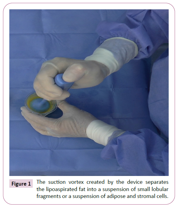
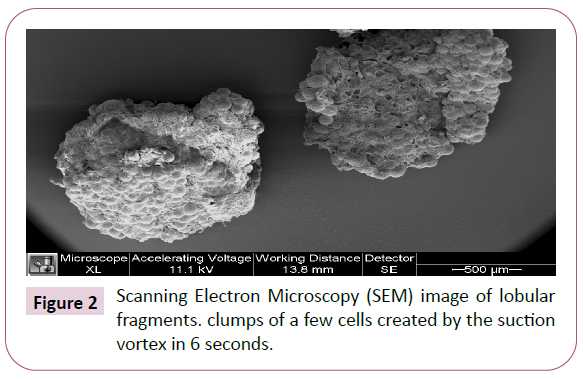
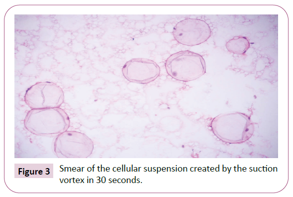
The subcutaneous fat in the human body is in an almost liquid state; for this reason, it can be aspirated through a cannula. As mentioned above, such techniques destroy biological material. To avoid these recent damaging methods, we aspirate the adipose tissue through a 4 mm diameter cannula with appropriately sized holes, in order to aspirate the original lobules of the tissue. In thin subjects, it is possible to use a 3 mm diameter cannula with holes proportional to the patient's adipose lobules. Cannulas with multiple holes reduce the suction force and damage the fat less. The lipoaspirate is then reduced by means of a suction vortex, which is created by a specially designed, economical, disposable, battery-driven, rotating device called an Adipopimer (Figure 1). The suction vortex reduces the size of the fat lobules in 5–6 seconds (Figure 2). In a further 20–30 seconds, it transforms the suspension of small lobular fragments into a suspension of living adipose and stromal cells (Figure 3).
Adipofilling
From 2001 to 2009, we were able to ascertain the efficacy of Adipofilling in the Department of Plastic and Reconstructive Surgery and Burns of San Martino Regional Hospital in Genoa, Italy. We named this technique "Adipofilling" to underline the importance of the adipocytes. The operations carried out in that period included the closure of an ulcer due to radiodermatitis with the exposure of three vertebrae [4] and the intradermal treatment of atrophic striae of the abdominal skin [5]. From 2009 to 2018; we designed the Adipopimer (Korpo srl Italy). During that period, we treated a rare case of bilateral facial atrophy (Figure 4) [6]. This patient required three Adipofilling sessions. The sequence of photos makes us understand the importance of the trophism of the receiving area.
Methodology
The phases of Adipofilling are: marking out the area of liposuction, local anaesthesia, liposuction, washing, preparation for lobular fragmentation and cell separation, supplementation with insulin, separation of the lipoaspirate by means of the Adipopimer, decantation or low-speed centrifugation to eliminate the separation liquids, transfer to 1-ml, 3-ml, and 5-ml Luer-lock syringes through a connecting tube, subdermal and subcutaneous injection through a cannula, intradermal injection with a needle, and preservation of the suspensions at temperatures from -20°C to -81°C.
Marking out the area of liposuction
Adipose tissue can be aspirated from many regions of the body. The lobular structure of the tissue may vary from one region to another, in terms of both size and its stromal component [7].
Local anaesthesia
The tumescent local anaesthetic is a modified Klein solution. For small quantities, we use 10 ml of 2% mepivacaine, 150 ml of lactate Ringer or physiological solution, and 1 mg of epinephrine. For larger quantities, we use 500 ml of lactate Ringer or saline solution, 30 ml of 2% mepivacaine, and 2 mg of epinephrine. Mepivacaine causes less damage to biological material than lidocaine does [8].
Liposuction
Liposuction is carried out through a 4-mm-diameter cannula. Large-calibre cannulas with large holes minimize the damage to the cells that is caused by friction and maintain the integrity of the lobules [9]. In thin subjects we use a 3-mm-diameter cannula. The holes of this cannula must also be proportional to the size of the fat lobules. Although small amounts of fat (e.g., 25 ml) can be aspirated and processed, we normally consider 50 ml to be the minimum quantity for Adipofilling of the face. The operator must consider the advantages of having a proper amount of material to inject and a reserve of material, which will be kept in a lockable freezer.
Washing in a flask
The lipoaspirate is placed in a flask with a tap, partially filled with lactate Ringer or saline solution. The washing liquid is drained off through the tap into sterile containers. Washing must continue until the liquid becomes clear and transparent; 2 litres of lactate Ringer or saline solution is normally enough to wash more than 200 ml of lipoaspirate. This method of washing is economical and quick. If a small amount of fat has been aspirated, it can be washed inside the same 60-ml catheter syringe used for liposuction. Despite thorough washing, a small quantity of anaesthetic solution remains in the lipoaspirate; this is often sufficient to achieve slight analgesia during the injection of the cellular suspension through the cannulas.
Preparation for lobular fragmentation and cell separation
Once washing has been completed, the lipoaspirate is poured into a beaker. A volume of lactate Ringer or saline solution three times greater than that of the fat must also be poured into the beaker. For the suction vortex to work properly, the biological material must have enough space to separate without being damaged.
Supplementation with insulin
To the separation liquid, we add 10–20 ml of 5% glucosate solution and 40–100 IU of ultra-rapid insulin, according to the amount of lipoaspirate to be processed. Insulin facilitates the entry of glucose and fatty acids into the adipocytes [10]. Insulin counters the lipolytic action of catecholamines [11] and plays a fundamental role in modulating the final phases of differentiation of the adipocytes. We also believe that insulin is useful in the cryo-preservation of the suspensions.
Separation of the lipoaspirate with the Adipopimer
The Adipopimer is equipped with a stabilized ceramic (Y-TZP) blade of 1/3 mm thickness, which rotates at 133.3 revolutions per second. In 1 or 2 seconds the zirconia blade separates the lobules. The lighter fragments are separated by the suction vortex created by the high-speed propeller of the disposable device. The operator immerses the "bell end" of the device into the lipoaspirate, then switches the device on and moves it up and down.
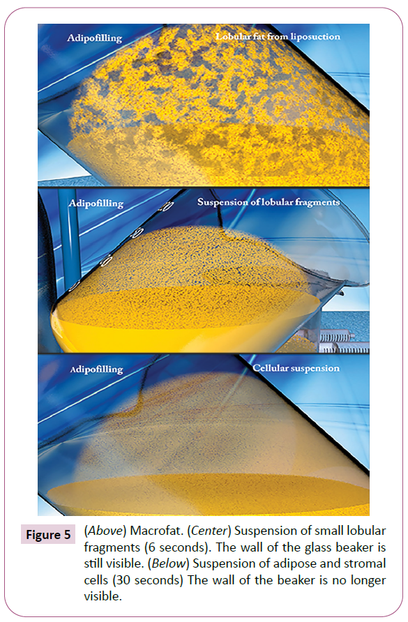
The lobules are reduced in size in about 6 or 7 seconds. The operator can check the particle size of the biological material by inclining the beaker. To create a suspension of single living cells, the operator activates the Adipopimer for another 20–25 seconds. On inclining the beaker, a uniform layer of cells will now be seen on the glass. This open system enables us to visually check the particle size obtained (Figure 5). One of the substances produced by the adipocyte is cathelicidin, a potent bactericidal agent [12]. This explains why we have never had any cases of infection in the hundreds of procedures that we have performed. The fact that the particles obtained with the suction vortex can be seen on the wall of the glass beaker is important. Indeed, fat differs from patient to patient and in the different sampling areas; it is therefore preferable to see what is being injected.
After separation, the total injectable volume is reduced by about 40%. In volumetric enhancement procedures, this reduction must be borne in mind during the liposuction phase. To the volumetric suspension of lobular fragments, we always add a small amount of cell suspension as supplementation, except in the case of breasts of women aged over 40 years. Single cells are free from contact inhibition and can freely secrete their products. Adipofilling involves minimal manipulation of biological material.
Decantation or low-speed centrifugation
The suspensions created by the Adipopimer are poured into 20-ml Luer-lock syringes from which the plungers have been removed. The syringes are capped and placed vertically in a beaker, where they are left to stand until the lighter organic material floats to the top of the separation liquid. After about 15 minutes, the cap is removed and, using a finger to control the flow, the operator drains off the liquid. The plunger is then reinserted, and the syringe is turned upside down. Another option is to centrifuge the cell suspension at 32 μg for 3 minutes, to eliminate the separation liquid and to concentrate the biological material. After centrifugation, the suspensions are poured into a beaker, which is delicately rotated to redistribute the stromal cells and the heavier adipocytes. This manoeuvre enables us to avoid having to pass the suspension several times through a connecting tube between two syringes, which would damage the living material.
Transfer to Luer-lock syringes
By means of a connecting tube, 1-ml, 3-ml, or 5-ml Luer-lock syringes are filled.
Subdermal and subcutaneous injection
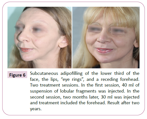
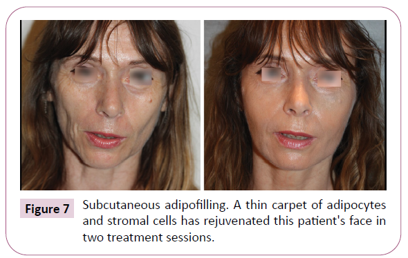
In the face, the suspensions are injected into the sites of volume deficit (Figure 6). The creation of a superficial "carpet" of adipose and stromal cells over the entire face also has a very pleasing effect (Figure 7).
Figure 6: Subcutaneous adipofilling of the lower third of the face, the lips, “eye rings”, and a receding forehead. Two treatment sessions. In the first session, 40 ml of suspension of lobular fragments was injected. In the second session, two months later, 30 ml was injected and treatment included the forehead. Result after two years.
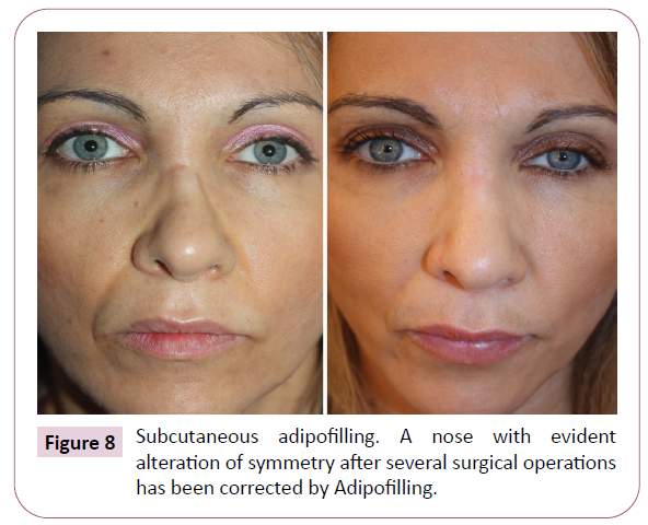
We graft adipose and stromal cells into the subcutaneous tissue, even immediately beneath the dermis. Superficial injection is physiological (fat with fat) and does not present complications. To correct noses that have undergone previous surgery (Figure 8), we use 25G needles where the cannula cannot penetrate. In the nose, the concentration of biological material is obtained by means of low-speed centrifugation.
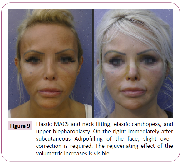
We often perform Adipofilling after elastic MACS and neck lifting. In this way, the correction of drooping facial volumes is optimized by volumetric and/or cellular restoration. During the same session, Adipofilling can be carried out in the temporal, malar, perioral and mandibular regions and in the cheeks and neck. This is possible because, in the elastic lifting procedures, we have eliminated the dissection of the cheeks and neck.
The intraoperative assessment of the volumetric result of Adipofilling is both visual and tactile. If we are satisfied with the visual result, we must, in any case, run a finger over the surface of the skin; wherever any area of diminished consistency of the tissues is detected, we inject additional cellular suspension. In the case of small corrections, the suspension of adipocytes and stromal cells may be more indicated, as it is more precise and controllable. Adipofilling of the face requires slight over-correction (Figure 9).
Intradermal injection
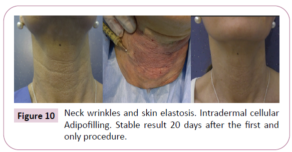
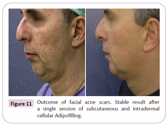
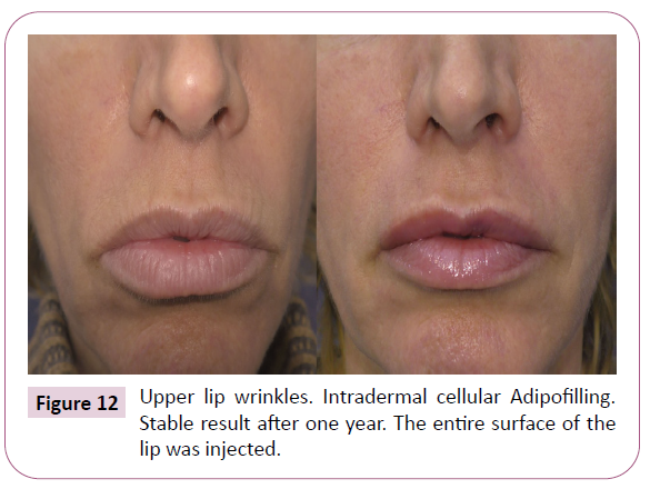
Injecting adipose and stromal cells into the dermis has no volumetric effect; it does, however, exert a very rapid and potent regenerative effect. When injected into the dermis, the adipose cells do not survive, but they have enough time to secrete the numerous substances that they produce; together with the stromal cells, they endow the tissues with new youth. Intradermal Adipofilling is quickly effective in elastosis (Figure 10), acne scars (Figure 11), lip wrinkles (Figure 12), etc. The intradermal suspension is injected with 21G needles. It is not advisable to use needles of smaller diameter, which traumatize the living material and make intradermal injection difficult.
Preservation of the suspensions at temperatures from -20°C to -81°C
Fat grafting normally requires multiple treatments and thus repeated liposuction to achieve treatment goals. The need for repeated liposuction limits the diffusion of the method. In the past, cryo-preservation at -80°C and -196°C, without the use of cryoprotective agents, yielded disappointing results [13].
Cryo-preservation with cryo-protectors such as bovine serum, dimethyl sulfoxide (DMSO), methylcellulose (MC) and/or polyvi nylpyrrolidone (PVP), sericin, etc. [14], is costly and difficult to implement in small clinics and, in terms of maintaining the greatest possible integrity of the adipocytes, is still unsatisfactory. Freezing damages the mitochondrial DNA [15].
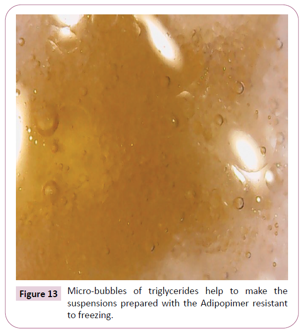
Biological material processed by means of the Adipopimer will not freeze. The suspensions can be conserved, without cryo-protectors, even for more than a year, in Luer-lock syringes, at temperatures from -20°C to -81°C. On removal from the freezer, this material can be injected immediately; it does not need to be thawed, as it is not frozen. The mechanisms whereby our suspensions do not freeze are still under investigation. First, lobular fragmentation and cellular separation break down the architecture of the tissue. The suspensions are therefore free from areas of water collection. Second, during liposuction and the processing of the lipoaspirate, small amounts of glycerol, phospholipids, and proteins are released. These substances remain among the cells even after washing. During processing, the oil separates into tiny droplets. The proteins are rapidly absorbed on the surface of these micro-bubbles, forming a membrane that covers them completely. This membrane stabilizes the micro-bubbles, preventing them from fusing. The micro-bubbles of triglycerides are surrounded by non-freezable water containing proteins, mucopolysaccharides and salts. The system is stable over time (Figure 13).
The third hypothesis is that the energy structures of the adipocytes receive, emit and accumulate energy in accordance with the principle of resonance. The Adipopimer is endowed with a special 1/3 mm blade made of stabilized ceramic (Y-TZP), which is known to be able to emit far infrared radiation (FIR) of 4–14 μm wavelengths. In both in vitro and in vivo studies this wavelength has been shown to stimulate living cells [16,17]. During the separation of the lipoaspirate, the ceramic structure (Y-TZP) pumps into the adipocytes the FIR biogenetic energy emitted by the ZrO2 quantum dots of the ceramic. Owing to the resonance effect, the altered waves of the adipocytes return to their normal frequency and recover the elastic property that was impaired by the mechanical stress exerted during liposuction. The FIR biogenetic energy is absorbed by the mitochondria, which, owing to resonance, oscillate at the same vibrational frequency as the ZrO2 quantum dots structured in the stabilized Y-TZP ceramic [18] in this way, they become resistant to freezing during cryo-preservation.
Results
The volumetric results of Adipofilling are clearly visible after even a single treatment. The cell suspension injected into the dermis rapidly regenerates aged and scarred skin. Shallow lip wrinkles, which do not need mixed peeling, also disappear. The cell suspension is particularly suitable for the rejuvenation of periorbital tissues. If it is injected in an excessive quantity, it can be re-aspirated with the same cannula (from 20 G to 25 G).
Discussion
Facial asymmetry, a volume deficit or an area of elastosis or wrinkles compromises the rejuvenating effect of an efficacious lifting procedure. Subcutaneous injection of the suspension of lobular fragments or intradermal injection of the suspension of single adipose and stromal cells has never caused any aesthetic complications; moreover, even after one single treatment, our patients have been satisfied with the persistence of the results achieved. In Adipofilling of the face, we believe that the minimum quantity of lipoaspirate should be 50 ml, which becomes 30 ml after volumetric reduction. Aspirating a greater quantity of fat enables some of it to be preserved for further corrections. The possibility of preserving the suspensions in a freezer, for more than a year, makes the procedure more economical. Only one session of liposuction enables us to carry out several sessions of Adipofilling. It must be borne in mind, however, that the quality of fat differs in different regions of the body. The fat in the buttock support areas is not good. By contrast, if the classic sampling areas cannot be exploited, the anterior region of the thighs can be used. Although Adipofilling is performed according to a standardized procedure, the variables are many and also concern the recipient area, age and the general condition of the patient.
Our goal was to reduce the particle size of the lipoaspirate, while maintaining the integrity of the adipocytes, in order to exploit both their volumetric contribution and the numerous substances that they produce: peptide hormones, paracrine, cytokines, adipsin, (ASP), exosomes, leptin, angiotensinogen, (PAI-1), adiponectin, resistin, cathelicidin, steroids hormones, and others.
Liposuction through a 4 mm cannula and subsequent volumetric reduction by means of the suction vortex results in the creation of a living material of small dimensions that can be grafted with success even into poorly vascularized tissues. The Adipopimer enables us to obtain very large quantities of suspensions of small fragments and single cells in a few minutes. This allows us to modify the face and neck as we please.
We not only correct small volumetric deficiencies; if necessary, we also modify the “structure“of the face. Before Adipofilling, if the skin at the sideburns can be pinched between two fingers, we perform an elastic MACS lift. If the skin on the sideburns cannot be pinched, we perform Adipofilling.
The regenerative efficacy of a suspension of adipose and stromal celle can best seen in aged skin that has elastosis or wrinkles; in such cases, the effects of intradermal injection are cleary visible, documentable, and traceable over time. Research in living tissue immediately achieves the goal: check the effectiveness of the procedures. The relationship between adipocytes and stromal cells must be maintained. After a prolonged actuation of the device and a prolonged low-speed centrifugation, stromal cells can be extracted.
If the adipocyte suspension is depleted of stromal cells when it is injected into the dermis, it will not have the same regenerative effect as the complete suspension. Likewise, injecting only the stromal portion, which is visible after centrifugation as a thin white layer below the adipocytes, does not produce the desired clinical results. Today the suspension of adipocytes and stromal cells together is the most powerful regenerative tool in our possession.
Conclusion
The Adipofilling suspensions constitute a versatile tool for correcting volume deficits, skin ageing, and scarred tissue. This technique enhances the value of adipose tissue. Fat is a precious, biologically active treasure; operators and patients alike should reflect on its possible immediate and future use.
References
- Coleman SR (2001) Structural fat grafts. Clin Plast Surg 28: 111-119.
- Ferraro GA, De Francesco F, Tirino V, Cataldo C, Rossano F, et al. (2011) Effects of a new centrifugation method on adipose cell viability for autologous fat grafting. Aesthetic Plast Surg 35: 341.
- Tonnard P, Verpaele A, Peeters G, Hamdi M, Cornelissen M, et al. (2013) Nanofat grafting: basic research and clinical applications. Plast Reconstr Surg 132: 1017-1026.
- Capurro S (2018) Chronic radiodermatitis with exposure of three vertebrae: treatment with Adipofilling R. CRPUB Med Video J Adipofilling section.
- Capurro S (2016) Treating stretch marks with Adipofilling. CRPUB Med Video J Adipofilling section.
- Capurro S (2017) Correction of severe bilateral facial atrophy by adipofilling. CRPUB Med Video J Adipofilling section.
- Esteve D, Boule N, Belles C, Zakaroff-Girard A, Decaunes P, et al. (2019) Lobular architecture of human adipose tissue defines the niche and fate of progenitor cells. Nat Commun 10.
- Keck M, Zeyda M, Gollinger K, Burjak S, Kamolz L, et al. (2010) Local anesthetics have a major impact on viability of preadipocytes and their differentiation into adipocytes. Plast Reconstr Surg 126: 1500-1515.
- Ozsoy Z, Kul Z, Bilir A (2006) The role of cannula diameter in improved adipocyte viability: a quantitative analysis. Aesthet Surg J 26: 287-289.
- Czech MP (2002) Fat targets for insulin signaling. Mol Cell 9: 695-6966.
- Lonnqvist F, Nyberg B, Wahrenberg H, Arner P (1990) Catecholamine-induced lipolysis in adipose tissue of the elderly. J Clin Invest 85: 1614-1621.
- Zhang LJ, Guerrero-Juarez CF, Hata T, Bapat SP, Ramos R, et al. (2015) Dermal adipocytes protect against invasive Staphylococcus aureus skin infection. Sci 347: 67-71.
- Mashiko T, Wu SH, Kanayama K, Asahi R, Shirado T, et al. (2018) Biological Properties and Therapeutic Value of Cryopreserved Fat Tissue. Plast Reconstr Surg 141: 104-115.
- Miyamoto Y, Oishi K, Yukawa H, Noguchi H, Sasaki M, et al. (2012) Cryopreservation of human adipose tissue derived stem/progenitor cells using the silk protein sericin. Cell Transplant 21: 617-622.
- Reardon AJF, Elliott JAW, McGann LE (2015) All Investigating membrane and mitochondrial cryobiological responses of HUVEC using interrupted cooling protocols. Cryobiology 71: 306-317.
- Chang JC, Wu SL, Hoel F, Cheng YS, Ko-Hung Liu, et al. (2016) Far-infrared radiation protects viability in a cell model of Spinocerebellar Ataxia by preventing poly-Q protein accumulation and improving mitochondrial function. Sci Rep 6: 30436.
- Vatanserver F, Hamblin MR (2012) Far infrared radiation (FIR): its biological effects and medical applications. Photonics Lasers Med 4: 255-266.
- Shirvanimoghaddam K, Khayyam H, Abdizadeh H, Akbari MK, Pakseresht AH, et al. (2016) Effect of B4C, TiB2 and ZrSiO4 ceramic particles on mechanical properties of aluminium matrix composites: Experimental investigation and predictive modelling. Ceram Int 42: 6206-6220.
Open Access Journals
- Aquaculture & Veterinary Science
- Chemistry & Chemical Sciences
- Clinical Sciences
- Engineering
- General Science
- Genetics & Molecular Biology
- Health Care & Nursing
- Immunology & Microbiology
- Materials Science
- Mathematics & Physics
- Medical Sciences
- Neurology & Psychiatry
- Oncology & Cancer Science
- Pharmaceutical Sciences
