Elastic Canthopexy
Ospedale Policlinico San Martino, Centro di ricerca medica, Genova Italia
- *Corresponding Author:
- Dr. Sergio Capurro
Ospedale Policlinico San Martino
Centro di ricerca medica
Via Rimassa 51/8 16129 Genova, Italy.
E-mail: sergio.capurro@hotmail.it
Received Date: September 19, 2021; Accepted Date: October 06, 2021; Published Date: October 13, 2021
Citation: Capurro S (2021) Elastic Canthopexy. J Aesthet Reconstr Surg Vol.7 No.7:42.
Abstract
Background: Operators and patients alike have always wanted a mini-invasive surgical procedure that is simple, safe and without complications – a procedure that can create the almond-shaped eyes of youth.
Methods: The external canthus and the tissues lateral to it are placed under traction by an Isosceles suspension triangle that is created by means of the elastic thread and the two-tipped needle (Elasticum, Korpo). The base of the triangle is situated at the hairline, while its apex is immediately below the lateral canthus.
Results: The result of elastic canthopexy is immediately visible; the patient's look is more youthful and attractive. This procedure can simply lengthen the eyes or else modify their inclination, creating the appearance of cats' eyes.
Conclusion: Elastic canthopexy is a simple procedure that preserves the eyelids and the lateral corner of the canthus, thereby avoiding the complications of traditional canthopexy and canthoplasty procedures.
https://bluecruiseturkey.co
https://bestbluecruises.com
https://marmarisboatcharter.com
https://bodrumboatcharter.com
https://fethiyeboatcharter.com
https://gocekboatcharter.com
https://ssplusyachting.com
Keywords
Canthopexy; Elastic suture
Introduction
A canthopexy procedure is required to correct slackness of the anatomical elements that maintain the eyelids in tension, to modify the inclination of the eyes, to lengthen rounded eyes after lower blepharoplasty with skin incision, and to correct ectropion, etc. Today, canthopexy can also be included among the lifting techniques for facial rejuvenation. Indeed, rejuvenating the eyes perfects rejuvenation of the face.
Operators and patients alike have always wanted a mini-invasive surgical procedure that is simple, safe and without complications – a procedure that can create the almond-shaped eyes of youth. Elastic canthopexy meets these requirements. This procedure has been made possible by the invention of an elastic thread [1] mounted on a two-tipped surgical needle Jano needle [2,3]. Within a few weeks, connective cells colonize the elastic thread, transforming it into a "ligament" that stabilizes the result.
Materials and Methods
The external canthus and the tissues lateral to it are placed under traction by an Isosceles suspension triangle that is created by means of the elastic thread and the two-tipped needle. By means of this new surgical thread (Elasticum, Korpo srl), which is made of silicone and sheathed in polyester, subcutaneous traction can be exerted and suspension carried out without dissecting the tissues. The base of the triangle is situated at the hairline, into temporal fascia, while its apex is immediately below the lateral canthus, into the retinaculum oculi.
Preoperative design
Using a dermographic pen, the operator draws a line in a fold of a crow's foot, in order to establish the inclination to give the eyes. An identical line is then drawn in the corresponding expression fold of the contralateral eye. The lines drawn in the expression folds facilitate the symmetry of the preoperative design. The lowest line of the Isosceles triangle starts at a point immediately below the canthus, at 10 mm from its apex, and extends to the hairline. The base of the Isosceles triangle is now drawn at the hairline; this line will be between 2.5 cm and 3.5 cm in length. An identical Isosceles triangle is then drawn in the vicinity of the contralateral eye. The probable pathway of the frontal branch of the facial nerve is marked out; a straight line is drawn which runs from the lowest portion of the earlobe and passes at a distance of ½ cm from the outer tip of the eyebrow (Figure 1).
Local anesthesia
Local anesthesia is carried out along the pathway where the elastic thread is to be implanted. To a 10 ml vial of 2% mepivacaine, ½ mg of epinephrine is added. After 10 minutes, the procedure begins.
Surgical procedure
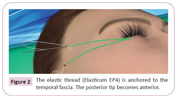
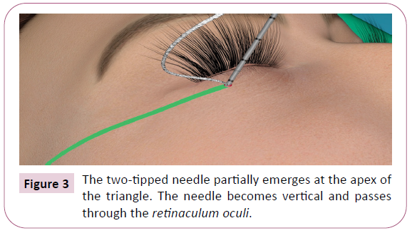
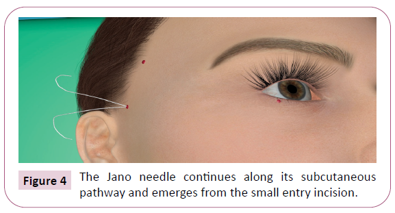
An incision of a few millimeters is made in the lower angle of the base of the triangle. Using a fine Klemmer, the operator creates a cavity to house the knot of the thread. The elastic thread (Elasticum EP4) is anchored to the temporal fascia by means of the two-tipped Jano needle. The two-tipped needle partially emerges, and the elastic thread is pulled through (Figure 2). A Klemmer is fixed to the end of the thread. The Jano needle is extracted until 5mm of the posterior tip remains in the tissues. The operator counts 5 and a half depth marks. At this point, the two-tipped needle is rotated, so that the posterior tip becomes anterior, and follows the direction of the previously drawn line, passing through the hypodermis above the superficial fascia. Once the Jano needle is beyond the frontal branch of the facial nerve, which has been marked in the preoperative design, it can penetrate more deeply and partially emerge at the preestablished point, below the canthus (Figure 3). The Jano needle is extracted until 5 mm of the tip remains in the tissues. It then rotates and passes obliquely in depth through the fibrous tissue of the retinaculum oculi. As the needle passes through the retinaculum, the operator feels greater resistance, which confirms that the elastic thread is properly anchored. On moving the needle, the eyelids must lengthen, and the palpebral fissure must narrow. The two-tipped needle continues along its pathway, emerging from the small entry incision (Figure 4).
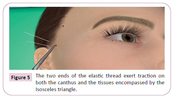
The elastic threads are pulled through, the eyelids lengthen, and the canthus is raised (Figure 5). After checking the result, the operator sutures the small incision. A small haemostatic wad of collagen may be inserted into the cavity before suturing, to maintain the knot in depth
Results
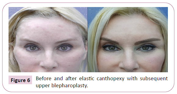
The bruising and swelling disappear within a few days; any slight alterations of the canthus disappear in less than three weeks. The result of elastic canthopexy is immediately visible; the patient's look is more youthful and attractive. This procedure can simply lengthen the eyes or else modify their inclination, creating the appearance of cats' eyes. In this latter case, the skin of the upper eyelid becomes slightly excessive; consequently, when the result has stabilized after a few months, upper blepharoplasty may be required (Figures 6 and 7). Lengthening the eyelids also corrects the appearance of eyes that are too close together.
The elastic thread does not cut into the tissues, is impalpable and invisible.
The results of elastic canthopexy remain stable over time. No complications have ever occurred in the 17 patients that we have operated on: 10 elastic canthopexy alone and 7 elastic canthopexy during elastic MACS lifting [3].
Discussion
In traditional canthopexy and canthoplasty, the procedures are carried out in the area of the eyelids and the canthus, without extending beyond the border of the orbit. The most common complication of these procedures is lower lid and canthal malposition due to the poor accuracy of primary fixation or subsequent loosening of the post-fixed canthus. Asymmetry and the loss of effective traction are frequent. Other common complications include deformities of the external commissure, recurrence, exposure of the mucosa and ectropion [4-6].
Moreover, when re-operating to correct these defects, the operator has to reopen the skin incisions and work on scar tissue.
In elastic canthopexy, there are no palpebral incisions and the canthal region remains undamaged. Elastic canthopexy exerts traction laterally or upward not only on the canthus but also on the tissues encompassed by the Isosceles triangle created by the elastic thread. It is as if the patient placed her fingertips at the sides of the eyes and, while pressing, moved her hands backwards and/or upwards. The procedure is easy; the only phase that requires particular attention is when the needle passes through the retinaculum.
The efficacy of elastic canthopexy is assessed during the procedure, while the patient is lying down; the operator judges how much the lateral portion of sclera has increased and, in this case, how the slope of the treated eye has been modified in comparison with the untreated contralateral eye.
Using a finger, the operator draws the external canthus of the treated eye laterally; there is no lengthening. If the same maneuver is performed in the contralateral region, which has not yet been treated, the canthus moves laterally. During the procedure, if the operator is not sure that the thread has been properly fixed to the retinaculum, he can immediately implant another thread along the same pathway as the previous one, which is not normally removed. Elastic canthopexy is able to correct the results not only of traditional canthopexy procedures, but also of previous elastic canthopexy if the patient is not completely satisfied with the effect achieved.
In this procedure, photographic documentation is important. In patients whose eyes are already elongated, the effect of elastic canthopexy will be chiefly preventive [7]. In these patients, it is advisable that the objective should be to visibly modify the inclination of the look.
Conclusion
Elastic canthopexy is a simple procedure that preserves the eyelids and the lateral corner of the canthus, thereby avoiding the complications of traditional canthopexy and canthoplasty procedures. Although it is a recent procedure, it has already proved efficacious in making the patient's look more appealing, rejuvenating over time the palpebral region, and correcting the appearance of hypotelorism.
References
- Capurro S (2004) Patent: Sheathed elastic surgigal thread. ITGE20030007 (A1): 7-28.
- Capurro S (1984) Un ago a due punte. Rivista Italiana di Chirurgia Plastica 16: 55-58.
- Capurro S (2016) Patent: Non-traumatic two-point needle for surgical suture. BRPI0610496 (A2): 11-16
- Capurro S (2021) Elastic canthopexy in elastic MACS and neck lifting. CRPUB Med Vid J.
- Kim YJ, Lee KH, Choi HL, Jeong EC (2016) Cosmetic Lateral Canthoplasty: Preserving the Lateral Canthal Angle. Arch Plast Surg 43: 316-320.
- Hester TR, Douglas T, Szczerba S (2009) Decreasing complications in lower lid and midface rejuvenation: The importance of orbital morphology, horizontal lower lid laxity, history of previous surgery, and minimizing trauma to the orbital septum: A critical review of 269 consecutive cases. Plast Reconstr Surg 123: 1037-1049.
- Oestreicher J, Mehta S (2012) Complications of Blepharoplasty: Prevention and Management. Plast Surg Int 2012: 1-10.
Open Access Journals
- Aquaculture & Veterinary Science
- Chemistry & Chemical Sciences
- Clinical Sciences
- Engineering
- General Science
- Genetics & Molecular Biology
- Health Care & Nursing
- Immunology & Microbiology
- Materials Science
- Mathematics & Physics
- Medical Sciences
- Neurology & Psychiatry
- Oncology & Cancer Science
- Pharmaceutical Sciences

