Elastic Mastopexy Maintains Glandular Integrity, Augments the Upper Quadrant, and Prevents Recurrence of Ptosis
Sergio Capurro*
Department of Plastic Surgery, Capurro Clinic, Genoa, Italy
Published Date: 2023-08-03DOI10.36648/2472-1905.9.2.159
Sergio Capurro*
Department of Plastic Surgery, Capurro Clinic, Genoa, Italy
- *Corresponding Author:
- Sergio Capurro
Department of Plastic Surgery,
Capurro Clinic, Genoa,
Italy,
E-mail: sergio.capurro@hotmail.it
Received date: July 03, 2023, Manuscript No. IPARS-23-17030; Editor assigned date: July 05, 2023, PreQC No. IPARS-23-17030 (PQ); Reviewed date: July 19, 2023, QC No. IPARS-23-17030; Revised date: July 26, 2023, Manuscript No. IPARS-23-17030 (R); Published date: August 03, 2023, DOI: 10.36648/2472-1905.9.2.159
Citation: Capurro S (2023) Elastic Mastopexy Maintains Glandular Integrity, Augments the Upper Quadrant, and Prevents Recurrence of Ptosis. J Aesthet Reconstr Surg Vol.9 No.2: 159.
Abstract
Background: Mastopexy is commonly defined as the removal of excess tissue, manipulation of the stroma and higher transposition of the nipple-areola complex. A breast implant is often inserted to project the upper quadrant. In reality, such operations would be more appropriately termed breast reduction and breast augmentation with skin removal. True mastopexy must keep the mammary stroma intact and never requires a prosthetic implant. The implantation of elastic threads (Elasticum EP4 Korpo SRL) that transform into a "ligament" prevents the recurrence of ptosis and enhances the upper quadrant.
Methods: The procedure involves de-epithelializing a shieldshaped area in the lower quadrant of the breast and reducing the diameter of the areola. The first elastic thread is implanted around the areola; if necessary, the apex of the breast is conized. Another elastic thread is implanted in order to create a suspensory ellipse. This thread runs from the anterior axillary pillar and through the subcutaneous tissue between the breast skin and the thoracic skin; it then passes at a slightly greater depth through the most prominent portion of the lower quadrant.
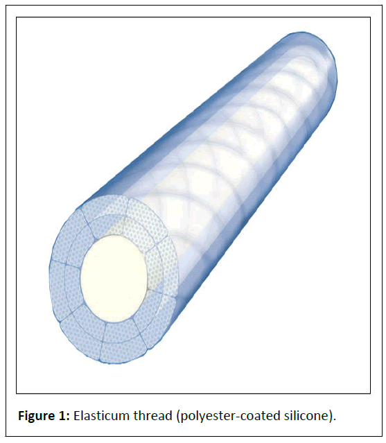
Results: Results were permanent. The breast is lifted, and the upper quadrant enlarged. This procedure significantly corrects breast asymmetry. If necessary we do an Adipofilling. The elasticum thread (polyester-coated silicone) is impalpable and does not cut into the tissues; when colonized by connective cells, it becomes a permanent "integrated ligament", making the correction of drooping stable.
Conclusion: Elastic mastopexy achieves stable esthetic modeling of the breasts, even heavy ones, under local anesthesia, while keeping the mammary stroma intact. The elastic suspension thread can be compared to a Cooper's ligament and is more resistant to the effects of gravity.
Keywords
Elastic mastopexy; Breast lift; Elastic suture; Elasticum thread; Two-tipped needle; Spolidoro mastopexy
Introduction
The common definition of mastopexy (Greek mastos "breast" + -pēxiā "affix") is as follows: “Mastopexy, or breast-lifting correction of sagging breasts, is a surgical procedure in which excess tissue (glandular, fatty, cutaneous) is removed, the stroma is manipulated to reduce tension on overstretched suspensory ligaments, excess skin is removed from the cutaneous envelope and the nipple-areola complex is repositioned higher up the mammary hemisphere. In surgical practice, mastopexy can be performed as a breast manipulation and reduction procedure or as an ancillary intervention within a combined mastopexy-breast augmentation procedure” [1,2]. This definition of mastopexy is incorrect. The above-mentioned operations are breast reduction and breast implant augmentation with skin removal, respectively. True mastopexy must keep the mammary stroma intact and never requires a prosthetic implant. To an exclusively cutaneous mastopexy with de-epithelialization (Spolidoro mastopexy) [3], we have added the implantation of elastic threads that prevent the areolae from dilating, conize the mammary apex and suspend the breasts, simultaneously preventing the recurrence of drooping and increasing the volume of the upper quadrant [4,5].
Materials and Methods
Elastic mastopexy involves de-epithelizing and suturing a shield-shaped area of the lower quadrant of the breast and raising the areola after periareolar de-epithelialization. A silicone-sheathed elastic thread [6] (Elasticum EP4 Korpo SRL) (Figure 1) mounted on a two-tipped Jano needle [7,8] is implanted around the areola to prevent it from dilating. If necessary, another elastic thread is implanted a few centimeters from the areola to conize the mammary apex. Liposuction through a 2.5 mm cannula removes the excess fat under the newly formed infra-mammary sulcus.
Through a hole made by a 16 G needle on the anterior axillary pillar, the two-tipped Jano needle is used to implant an elastic thread along an elliptical pathway in the subcutaneous tissue. After crossing the transition line between the breast skin and the thoracic skin in the upper quadrant and the most prominent line of the lower quadrant, the thread ascends and exits through the access hole. This thread permanently lifts the breast and augments the upper pole.
Preoperative design
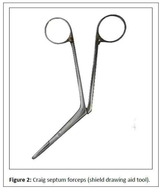
While the patient is standing, the operator marks out the infra-mammary sulci and the line that descends from the jugular towards the navel. Two points are marked out laterally at 3 cm from the jugular. These are the starting points of the straight lines that pass over the nipple and stop at the infra-mammary sulcus. On these lines, points are marked 18 cm, 20 cm and 22 cm from the jugular. To establish the width of the shield-shaped area, we press the end of the nasal forceps (Figure 2) on the line drawn under the areola and pinch the skin with two fingers to simulate what the new breast will look like. The operator's assistant uses a demographic pen to mark the width of the area to be de-epithelialized. This area, which extends downward to 1 cm above the infra-mammary sulcus and upward to the lower half of the areola, is then drawn. The operator lifts the breast with a hand and marks the line of transition between the breast skin and thoracic skin.
Local anesthesia
A dilute anesthetic solution (1 vial of 2% mepivacaine and 1 vial of 2% lidocaine in 250 ml lactate ringer's solution or saline+1 mg of adrenaline) is injected into the shield-shaped area and around the areola using a 4 cm 27 G needle and a 10 ml syringe.
Surgical procedure
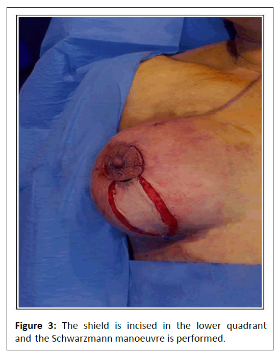
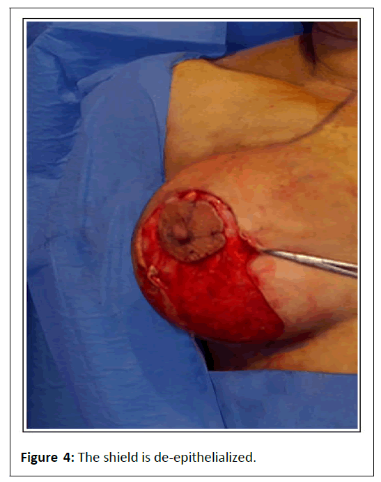
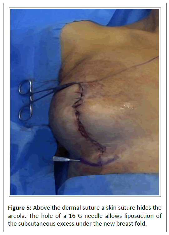
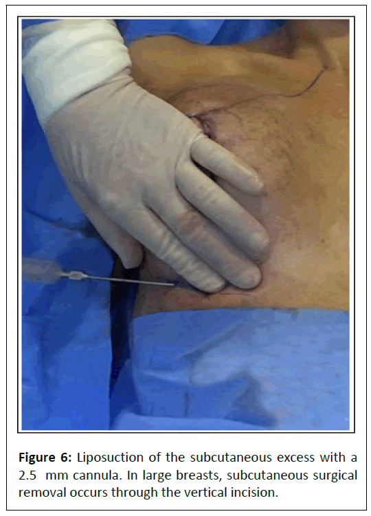
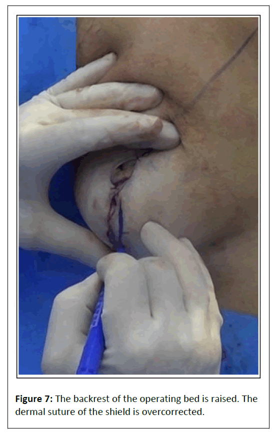
After administration of local anesthesia and the placement of reference stitches, de-epithelialization of the shield-shaped area is performed in the lower quadrant and around the areola (Schwarzmann maneuver) (Figure 3). Once the area has been de-epithelialized (Figure 4), the first dermal spiral suture is performed, which stops a few centimeters from the areola. The 4-0 absorbable suture has duration of 90 days. Separate cutaneous stitches are inserted to hide the areola. The vertical suture is completed with dermal stitches that persist for 30 days. A hole is made with a 16 G needle and dilated with the tip of a thin Klemmer. Liposuction of the excess fat under the newly formed breast sulcus is now carried out (Figures 5 and 6). The patient is placed in a sitting position by raising the back-rest of the operating bed to determine whether the lower quadrant needs further de-epithelialization in order to achieve the desired shape. Any areas requiring further de-epithelialization are marked with the dermographic pen. The result should be a slight curvature of the lower quadrant (Figure 7).
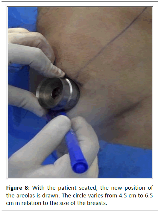
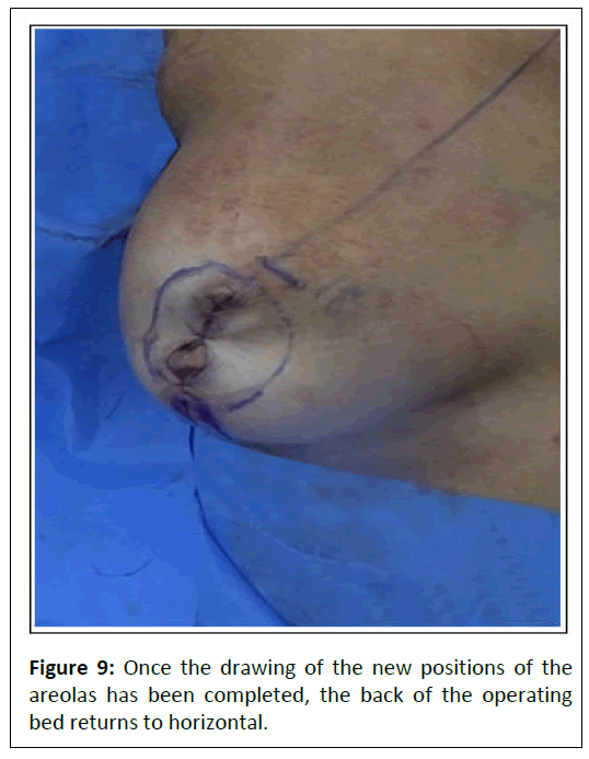
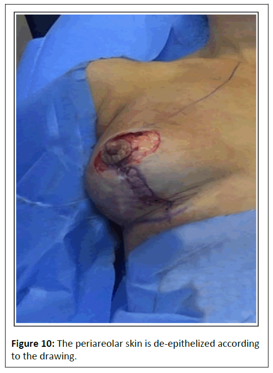
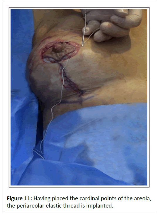
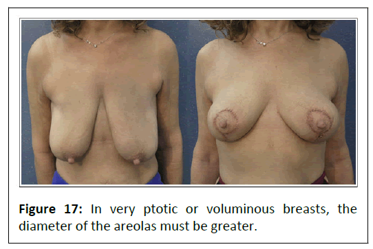
Overcorrection helps to maintain the result over time. Next, the position of the areolae is drawn. Circles ranging from 4.5 cm to 6.5 cm in diameter are used, depending on the amount of excess skin (Figure 8). If the breast is large and very ptotic, the diameter increases. Once the drawing of the new positions of the areolas has been completed, the back of the operating bed returns to horizontal (Figure 9). The periareolar skin is deepithelialized (Figure 10). The cardinal stitches of the areola are inserted. Before the areolar suture is completed, an elastic thread is implanted ½ cm from the areola to prevent it from dilating (Figure 11). The elastic thread is impalpable. If necessary, the operator conizes the apex of the breast by implanting a subcutaneous thread through a tunnel that encircles the areola at a distance of a few centimeters. The vertical suture is completed by means of rapid absorbable cutaneous stitches. The vertical suture of the de-epithelialized shield lifts and conizes the breast. Periareolar deepithelialization compresses the breast against the chest and helps to obtain a compact rounded shape.
Suspensory ellipse
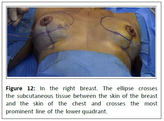
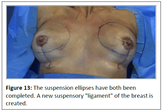
The operator lifts the breast with a hand and marks the line of transition between the breast skin and the thoracic skin. The most prominent line of the lower quadrant is now marked. This line joins the demarcation line between the breast skin and thoracic skin, completing the ellipse. The elastic thread that creates the suspensory ellipse is now implanted. Local anesthesia is performed along the part of the elliptical pathway that has not yet been anesthetized. A hole is made with a 16 G needle on the line of the ellipse at the level of the anterior axillary pillar. The hole is dilated and deepened with the tip of a thin Klemmer. The two-tipped Jano needle on which the elastic thread is mounted is inserted into the hole and rotated to follow the upper pathway at a depth of ½ cm to 1 cm. The needle partially emerges up to the desired depth, e.g., up to the 5th depth mark, and then rotates and continues along the pre-established pathway at a constant depth. To check that the depth is correct, the operator moves the Jano needle up and down; these movements must not give rise to skin introflections. In the lower half of the ellipse, the thread is implanted at a depth of 1 cm, or slightly more. The two-tipped needle exits through the access hole and the two ends of the elastic thread are placed under traction and knotted. With a Klemmer, the operator pushes the knot to the bottom of the skin hole, which is not sutured (Figures 12 and 13). An antibiotic ointment is applied to the vertical and periareolar suture, along the pathway of the threads, and to the access hole of the elastic thread of the suspensory ellipse.
Results
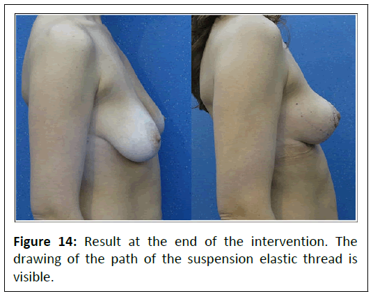
The result is immediately visible (Figure 14). The breast is stabilized, and the upper pole increased in volume. After elastic mastopexy, the patient can resume without delay, her normal activities.
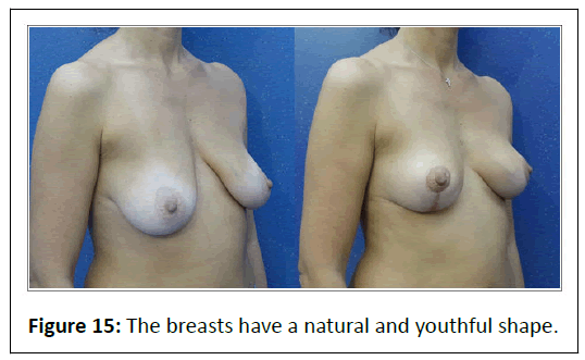
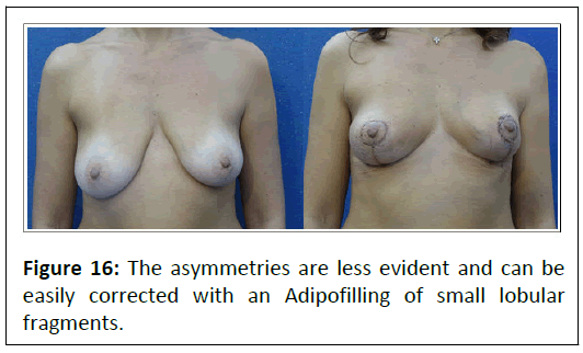
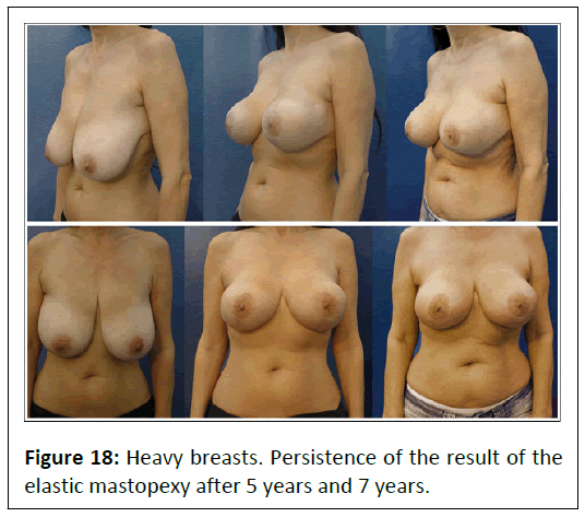
In the 23 patients we have treated, elastic mastopexy has yielded excellent natural results (Figure 15). The asymmetries are less evident (Figure 16) and can be easily corrected with an Adipofilling of small lobular fragments. In very ptotic breasts, the elastic mastopexy allows to obtain a pleasant shape (Figure 17). Even in those with heavy breasts implanting elastic threads and maintaining the integrity of the mammary stroma ensure that the result will persist over time (Figure 18).
Discussion
The most common complication after mastopexy is recurrent ptosis. Most surgeons believe that using skin as a “bra” is a recipe for failure. They also think that the correction of ptosis should be glandular and that it can be achieved by means of parenchymal reshaping. Glandular or dermo-glandular flaps with a superior [9-19], medial [20-22], supero-media [23-26], lateral [27-30], superior-lateral [31], or inferior [32-35] peduncle, bipedicled flaps [36,37], or auto-prostheses, as well as all other manipulative techniques that have been proposed, can yield good immediate results. In our experience, however, trophic damage inevitably leads to reduction of the glandular volume and, consequently, the recurrence of ptosis after less than 2 years. Therefore, the patient is left with smaller drooping breasts and a deficient upper pole. Even periosteal and fascial fixations to the chest wall [38], in addition to being non-physiological, do not guarantee persistent results. Due to these procedures, which do not respect the anatomy and biology of the tissues, operators are often forced to insert breast implants, even in patients with breasts of adequate volume. We avoid techniques that impair the trophism of the breast tissue.
It is preferable to perform an exclusively cutaneous procedure with de-epithelialization, which has the advantage of maintaining glandular integrity and correcting ptosis without loss of volume. Of course, overcorrection is necessary; de-epithelialization of the shield-shaped area in the lower quadrant must consider the natural sagging of the skin.
In mastopexy procedures, it is also necessary to take oncological aspects into account. Most breast tumors occur in patients with high stromal density [39]. Such patients need to be followed up more carefully, and unmanipulated breast tissue facilitates precise and early diagnosis.
Elastic mastopexy now enables us to obtain a natural, youthful breast without volume deficits in the upper pole and without recurrence of ptosis. The solution that we propose is inspired by anatomical studies. The breast is supported by fibrous septa composed of connective tissue, which support the internal adipose tissue and mammary gland (Cooper's ligament) [40,41]. Our elastic suspension thread replaces Cooper's ligament, lifting the mammary gland and reducing its weight on the skin, and has the advantage of being permanent. The elastic suspension thread alone can prevent breast ptosis in young women with particularly dense and heavy breast stroma.
Spolidoro elastic mastopexy is an updated procedure that can be adapted to any type of sagging breast and has the advantage of not requiring dissections or glandular flaps, which damage the trophism of the tissues. Vascular damage inevitably causes reduction in breast volume and recurrence of ptosis. This procedure significantly corrects breast asymmetry. However, if further corrections are needed, we perform Adipofilling, which utilizes small lobular fragments [42,43].
Moreover, the elastic threads that conize and suspend the breast can correct the shape and ptosis of breasts with implants. In this case, access is through an 8 mm periareolar incision, and a hole made with a 16 G needle. In prosthetic breasts, the elastic threads are implanted at a depth of ½ cm [44,45].
Conclusion
With a simple operation under local anesthesia, elastic mastopexy solves the problems of traditional operations with manipulation of the glandular tissues: Use of general anesthesia, loss of volume, deficiency of the upper pole, recurrence of ptosis, the non-juvenile form, dificulties in instrumental examinations, and the possibility of having to place an implant to correct the shape and volume of the breast. A minimally invasive procedure such as Spolidoro mastopexy combined with a new technology that involves an elastic surgical thread equipped with a two-tipped needle that imitates the suspensory ligaments of the breast allows high-quality and long-lasting natural results to be obtained.
References
- De la TJ (2009) Breast mastopexy. Medscape.
- Anastasatos JM (2010) Medial pedicle and mastopexy breast reduction. Medscape.
- Capurro S (2010) Vertical spolidoro's mastopexy. CRPUB medical video journal. Elastic plastic surgery section.
- Capurro S, Berlanda M (2016) Using the elastic thread in aesthetic breast surgery. CRPUB medical video journal. Elastic plastic surgery section.
- Capurro S (2017) Conization of breasts that have silicone implants by means of the elastic thread and Jano needle. CRPUB medical video journal. Elastic plastic surgery section.
- Capurro S (2004) Sheathed elastic surgical thread. ITGE20030007 (A1): 7-28.
- Capurro S (1984) Un ago a due punte. Rivista Italiana di Chirurgia Plastica 16: 55-58.
- Capurro S (2016) Patent: Non-traumatic two-point needle for surgical suture. BRPI0610496 (A2): 11-16.
- Pitanguy I (1967) Surgical correction of breast hypertrophy. Br J Plast Surg 20: 78-85.
- Lassus C (1970) A technique for breast reduction. Int Surg 531: 69-72.
[Indexed]
- Weiner DL, Aiache AE, Silver L, Tittiranonda T (1973) A single dermal pedicle for nipple transposition in subcutaneous mastectomy, reduction mammaplasty, or mastopexy. Plast Reconstr Surg 512: 115-120.
[Crossref], [Google Scholar], [Indexed]
- Arufe HN, Erenfryd A, Saubidet M (1977) Mammaplasty with a single, vertical, superiorly-based pedicle to support the nipple-areola. Plast Reconstr Surg 602: 221-227.
[Crossref], [Google Scholar], [Indexed]
- Hugo NE, McClellan RM (1979) Reduction mammaplasty with a single superiorly based pedicle. Plast Reconstr Surg 632: 230-234.
[Crossref], [Google Scholar], [Indexed]
- Marchac D, de Olarte G (1982) Reduction mammaplasty and correction of ptosis with a short inframammary scar. Plast Reconstr Surg 691: 45-55.
[Google Scholar], [Indexed]
- Elsahy NI (1982) The hexagonal technique for mastopexy and reduction mammaplasty. Aesthet Plast Surg 62: 107-115.
[Crossref], [Google Scholar], [Indexed]
- Finger RE, Vasquez B, Drew GS, Given KS (1989) Superomedial pedicle technique of reduction mammoplasty. Plast Reconstr Surg 833: 471-478.
[Crossref], [Google Scholar], [Indexed]
- Lejour M (1994) Vertical mammaplasty and liposuction of the breast. Plast Reconstr Surg 941: 100-114.
[Crossref], [Google Scholar], [Indexed]
- Lassus C (1996) A 30-year experience with vertical mammaplasty. Plast Reconstr Surg 972: 373-380.
[Crossref], [Google Scholar], [Indexed]
- Ribeiro L, Accorsi A, Buss A, Marcal-Pessoa M (2002) Creation and evolution of 30 years of the inferior pedicle in reduction mammaplasties. Plast Reconstr Surg 1103: 960-970.
[Crossref], [Google Scholar], [Indexed]
- Graf R, Biggs T (2002) In search of better shape in mastopexy and reduction mammaplasty. Plast Reconstr Surg 1101: 309-317.
[Crossref], [Google Scholar], [Indexed]
- Asplund OA, Davies DM (1996) Vertical scar breast reduction with medial flap or glandular transposition of the nipple-areola. Br J Plast Surg 498: 507-514.
[Crossref], [Google Scholar], [Indexed]
- Nahabedian MY, McGibbon BM, Manson PN (2000) Medial pedicle reduction mammaplasty for severe mammary hypertrophy. Plast Reconstr Surg 1053: 896-904.
[Crossref], [Google Scholar], [Indexed]
- Abramson DL, Pap S, Shifteh S, Glasberg SB (2005) Improving long-term breast shape with the medial pedicle wise pattern reduction. Plast Recontr Surg 1157: 1937-1943.
[Crossref], [Google Scholar], [Indexed]
- Orlando JC, Guthrie RH (1975) The superomedial dermal pedicle for nipple transposition. Br J Plast Surg 281: 42-45.
[Crossref], [Google Scholar], [Indexed]
- Hauben DJ (1984) Experience and refinements with the superomedial dermal pedicle for nipple-areola transposition in reduction mammaplasty. Aesthet Plast Surg 83: 189-194.
[Crossref], [Google Scholar], [Indexed]
- Hall-Findlay EJ (1999) A simplified vertical reduction mammaplasty: Shortening the learning curve. Plast Reconstr Surg 1043: 748-759.
[Crossref], [Google Scholar], [Indexed]
- Spear LS, Davison SP, Ducic I (2004) Superomedial pedicle reduction with short scar. Semin Plast Surg 183: 203-210.
[Crossref], [Google Scholar], [Indexed]
- Skoog T (1963) A technique of breast reduction: Transposition of the nipple on a cutaneous vascular pedicle. Acta Chir Scand 126: 453-465.
[Google Scholar], [Indexed]
- Nicolle F (1982) Improved standards in reduction mammaplasty and mastopexy. Plast Reconstr Surg 693: 453-459.
[Crossref], [Google Scholar], [Indexed]
- Botta SA, Rifai R (1991) Personal refinements in the single pedicle skoog technique for reduction mammaplasty. Aesthet Plast Surg 153: 257-264.
[Crossref], [Google Scholar], [Indexed]
- Blondeel PN, Hamdi M, Van de Sijpe KA, van Landuyt KH, Thiessen FE, et al. (2003) The laterocentral glandular pedicle technique for breast reduction. Br J Plast Surg 564: 348-359.
[Crossref], [Google Scholar], [Indexed]
- Strauch B, Elkowitz M, Baum T, Herman C (2005) Superolateral pedicle for breast surgery: An operation for all reasons. Plast Reconstr Surg 1155: 1269-1277.
[Crossref], [Google Scholar], [Indexed]
- Ribeiro L (1975) A new technique for reduction mammaplasty. Plast Reconstr Surg 553: 330-334.
[Crossref], [Google Scholar], [Indexed]
- Robbins TH (1977) A reduction mammaplasty with the areola-nipple based on an inferior dermal pedicle. Plast Reconstr Surg 591: 64-67.
[Crossref], [Google Scholar], [Indexed]
- Courtiss EH, Goldwyn RM (1977) Reduction mammaplasty by the inferior pedicle technique. An alternative to free nipple and areola grafting for severe macromastia or extreme ptosis. Plast Reconstr Surg 594: 500-507.
[Google Scholar], [Indexed]
- Hammond DC (1999) Short Scar Periareolar-Inferior Pedicle Reduction (SPAIR) mammaplasty. Plast Reconstr Surg 1033: 890-901.
[Crossref], [Google Scholar], [Indexed]
- Strömbeck JO (1960) Mammaplasty: Report of a new technique based on the two-pedicle procedure. Br J Plast Surg 13: 79-90.
[Crossref], [Google Scholar], [Indexed]
- McKissock PK (1972) Reduction mammoplasty with a vertical dermal flap. Plast Reconstr Surg 493: 245-252.
[Crossref], [Google Scholar], [Indexed]
- Ritz M, Silfen R, Southwick G (2006) Fascial suspension mastopexy. Plast Reconstr Surg 1171: 86-94.
[Crossref], [Google Scholar], [Indexed]
- Fernández-Nogueira P, Mancino M, Fuster G, Bragado P, Puig MP, et al. (2020) Breast mammographic density: Stromal implications on breast cancer detection and therapy. J Clin Med 9: 776.
[Crossref], [Google Scholar], [Indexed]
- Rehnke RD, Groening RM, Van Buskirk ER, Clarke JM (2018) Anatomy of the superficial fascia system of the breast: A comprehensive theory of breast fascial anatomy. Plast Reconstr Surg 142: 1135-1144.
- Capurro S (2008) Breast enhancement by means of a suspension of adipose and stromal cells: Adipofilling®. CRPUB medical video journal. Adipofilling section.
- Capurro S (2022) Elastic "round block" and adipofilling of "hollowed out" breasts, without skin excision. CRPUB medical video journal. Elastic plastic surgery section.
- Capurro S (2019) Conization and mastopexy of breasts with silicone implants by means of two incisions of a few millimeters and the elasticum thread and Jano needle. CRPUB medical video journal. Elastic plastic surgery section.
- Capurro S (2022) Elastic conization and suspension improves the appearance of breasts with implants. J Aesthet Reconstr Surg 8: 1-4.
Open Access Journals
- Aquaculture & Veterinary Science
- Chemistry & Chemical Sciences
- Clinical Sciences
- Engineering
- General Science
- Genetics & Molecular Biology
- Health Care & Nursing
- Immunology & Microbiology
- Materials Science
- Mathematics & Physics
- Medical Sciences
- Neurology & Psychiatry
- Oncology & Cancer Science
- Pharmaceutical Sciences
