Naso-Orbital Complex Reconstruction with Titanium Mesh and Canthopexy
María Genma Pérez Paredes, Alicia Pérez Bustillo, Beatriz González Sixto, Manuel Ángel and Rodríguez Prieto
DOI10.4172/2472-1905.100011
María Genma Pérez Paredes*, Alicia Pérez Bustillo, Beatriz González Sixto, Manuel Ángel and Rodríguez Prieto
Asistential Universitary complex of León, Spain
- *Corresponding Author:
- María Genma Pérez Paredes
Asistential Universitary complex of León, Spain.
Tel: 607540717
E-mail:pegemina@yahoo.es
Received date: December 12, 2015; Accepted date: January 02, 2016; Published date:January 12, 2016
Citation: Paredes MGM, Bustillo AP, Sixto BG, et al. Naso-Orbital Complex Reconstruction with Titanium Mesh and Canthopexy. J Aesthet Reconstr Surg. 2016, 2:2. doi: 10.4172/2472-1905.100011
Abstract
Context: We are introducing the reconstruction of two defects: squamous cell carcinoma located on the dorsum and right nasal sidewall, and sclerodermiform basal-cell carcinoma in the nasolabial fold and the nasal ala on the same side.
Case report: After removing the first tumor, we reconstructed the bone with a titanium mesh. We joined the two tarsi anchoring to the titanium mesh in order to create the internal canthal ligament. The second defect was reconstructed in different levels. For the nasal lining reconstruction, we designed a transposition flap over the nasolabial fold; leaving a pedicle that supplied nutrients to the flap which was turned 180º. The skin defect was reconstructed with a pedicled transpositional flap taken from the healthy skin betwaeen the two defects. The resulting skin defect was replaced by a paramedian forehead flap. The titanium mesh supplies a support for the neo-canthal ligament that provides an adequate opening of the eye lid and to avoid visual dysfunction and canthal dystopia.
Conclusion: The titanium mesh is a malleable system that provides an adequate support if there is lack of bone support. When the nasal defect is small but fullthickness, the cartilage substitution is not needed, because the used nasolabial and the paramedian forehead flaps provided enough consistency.
Keywords
Titanium mesh; Canthopexy; Naso-orbital reconstruction
Introduction
The defects resulting from the removal tumoral lesions may be located in different facial subunits and affecting different anatomical levels. We are presenting a complex naso-orbital reconstruction with titanium mesh and canthopexy.
Case Report
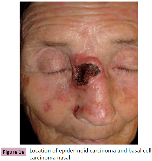
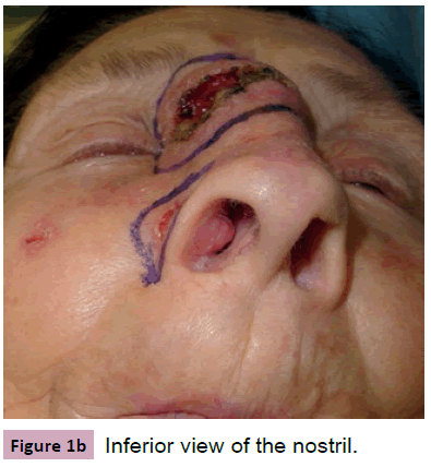
A 91 year old woman presenting a 2.7 cm by 2.5 cm squamous cell carcinoma located on the dorsum and right nasal sidewall. She also presented a sclerodermiform basal-cell carcinoma in the nasolabial fold and the nasal ala on the same side (Figure 1A and 1B).
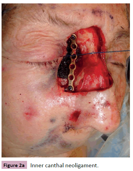
After removing the first tumor we reconstructed the bone with a titanium mesh fixed with screws to the internal supraorbital rim and the upper maxillar bone. We joined the two tarsi with 4-0 Nylon anchoring the inner end to the titanium mesh at the same level as the contralateral lacrimal crest (Figure 2A) to create the internal canthal ligament.
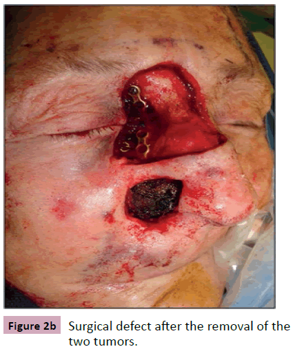
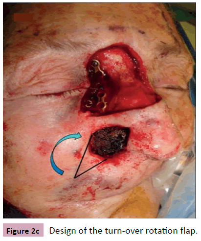
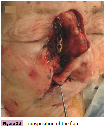
The second defect was reconstructed in different levels (Figure 2B). For the nasal lining reconstruction, we designed a transposition flap over the nasolabial fold; leaving a pedicle that supplied nutrients to the flap which was turned 180º, so the inner lining was created with a skin flap (Figure 2C). The borders of the nasal defect and the subcutaneous plane from the donor area were sutured with polyglycolic acid 4/0. The skin defect was reconstructed with a pedicled transpositional flap taken from the healthy skin between the two defects (Figure 2D).
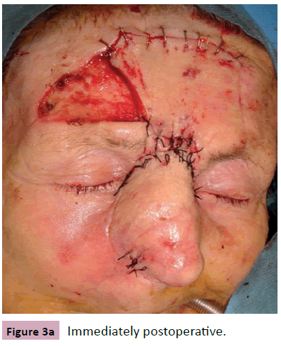
Finally, the resulting skin defect was replaced by a paramedian forehead flap, centered on the supratrochlear and supraorbital arteries (Figure 3A). The secondary frontal defect was covered with silicone dressing until the next surgery.
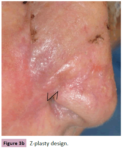
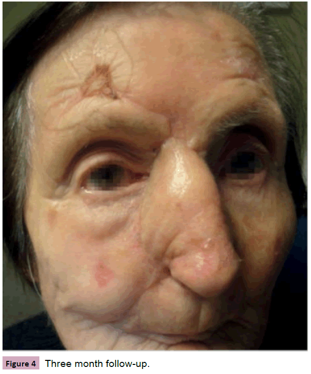
Three weeks later, the pedicle of the paramedian forehead flap was sectioned, the skin was readjusted to the nose defect and the excess skin was returned to its initial position in the frontal area leaving a small defect which healed by secondary intent. We did a Z-plasty to repair a minor defect of the alar rim (Figure 3B). After a 14 months follow-up there has been no relapse (Figure 4).
Discussion
In the first defect, the lack of bone support required the creation of a base for the posterior insertion of the canthal ligament, needed for the adequate opening of the eye lid and to avoid visual dysfunction and canthal dystopia. The titanium mesh is a malleable system that provides an adequate support and unlike the piton fixation and the transnasal wiring [1], is easy to place. The titanium mesh is also used for complex facial trauma reconstruction when there is lack of bone support. Its drawbacks are: infections, interference with radiological images and the need to adjust the radiotherapy dosimeter [2]. When we have bone support for the reconstruction and anchoring of the canthal ligament we can use the Mitek anchor system® [1].
When the nasal defect is full-thickness but of small size cartilage, substitution is not needed because the nasolabial and the paramedian forehead [3] flaps that we used, provided enough consistency [4], avoiding the collapse of the nostrils and the aesthetical alteration [5].
References
- Antonyshyn OM, Weinberg MJ, Dagmon AB (1996) Use of a new anchoringdevicefortendonreinsertion in medial canthopexy. PlastReconstrSurg 98: 520-523.
- Rodríguez-Prieto MA, Alonso-Alonso T, Sánchez- Sambucety P (2009) Nasal reconstructionwithtitaniummesh. DermatolSurg 35:282-286.
- Schreiber NT, Mobley SR (2011)Elegantsolutionsforcomplexparamedianforeheadflapreconstruction. Facial PlastSurgClinNorth Am 19:465-479.
- Lunatschek C, Schwipper V, Scheithauer M (2011)Softtissuereconstruction of thenose. Facial PlastSurg27: 249-257.
- Ulug BT, Kuran I (2008) Nasal reconstructionbasedonsubunitprinciplecombinedwithturn-overisland nasal skinflapfor nasal liningrestoration. Ann PlastSurg 61: 521-526.
Open Access Journals
- Aquaculture & Veterinary Science
- Chemistry & Chemical Sciences
- Clinical Sciences
- Engineering
- General Science
- Genetics & Molecular Biology
- Health Care & Nursing
- Immunology & Microbiology
- Materials Science
- Mathematics & Physics
- Medical Sciences
- Neurology & Psychiatry
- Oncology & Cancer Science
- Pharmaceutical Sciences
