Giant Ameloblastoma of the Mandible: An Exceptional Case Reports in the Early Childhood
Aloise Sagna, Aissata Ly and Ibrahima Fall
DOI10.4172/2472-1905.100017
Pediatric Surgery Department, Children Hospital Albert Royer, Dakar, Senegal
- *Corresponding Author:
- Aloise Sagna,Ph.D
Pediatric Surgery Department, Children Hospital Albert Royer
Chnear, Dakar, Senegal.
Tel: +221 3 3869 1818
E-mail: alosagna@hotmail.com
Received date: March 02, 2016; Accepted date: April 05, 2016; Published date: April 15, 2016
Citation: Sagna A, Ly A, FalI l. Giant Ameloblastoma of the Mandible: An Exceptional Case Reports in the Early Childhood. J Aesthet Reconstr Surg. 2016, 2:8. doi: 10.4172/2472-1905.100017
Abstract
Ameloblastoma is an odontogenic epithelium tumor very rare in children. It is histologically benign with locally extensive potential justifying the nickname “benign tumor with local malignancy”. The aim of our study was to start from a clinical case data and tried to bring out key elements of a specific therapeutic approach in children. We report the observation of a 5 y old girl, weighing 16 kg with a height of 1.05 m and presenting a voluminous tumor of the left mandible. The history taking noted the up raising marked by a progressive painless increase in size of the mandible without dental malocclusion, five months earlier. In her background check we noticed complete vaccination, heterozygote sickle cell disease at her 59 y old father. The examination found asymmetry of the lower part of the face and a voluminous mass blowing the alveolar rampart of the left mandible. The medical imaging evoked the diagnosis and histology after excision confirmed ameloblastoma. Then, reconstruction of the loss of bone by an inferior pedicled Facial Artery Muscular Mucosal (FAMM) flap was performed in the 24th postoperative day. There wasn’t distant recurrence of the tumor after 4 y follow up. The clinical feature is the age of our patient, the giant aspect of the tumor at the diagnosis time and the recourse to the FAMM flap for filling bone loss.
Keywords
Giant ameloblastoma; Tumour excision; Reconstruction; FAMM flap
Introduction
Ameloblastoma is an odontogenic epithelium tumour very rare in children. It is histologically benign with locally extensive potential justifying the nickname ‘‘benign tumour with local malignancy’’ [1]. Its early clinical diagnosis remains very difficult because of an asymptomatic long run and surgery is the only treatment although risk of reoccurrence and low rate of malignant degeneration [2]. The aim of our study was to highlight management difficulties from a 5 y old female clinical case data and bring out the elements of a child-specific therapeutic approach.
Case Report
We report the observation of a 5 y old girl of whom history taking noted disease up raising five months earlier marked by a progressive painless increase of the mandible without dental malocclusion. She was referred to us after consultation in departments of Stomatology and Paediatrics. In her background check we noticed complete vaccination, heterozygote sickle cell disease at her 59 y old father and tonsillectomy at her 39 y old mother.
Physical examination in March 17th, 2011 found a good condition with 16 kg of weight and a height of 1.05 m. There was an isolated voluminous mass blowing the alveolar rampart of the left mandible, lying down the symphysis and causing asymmetry of the lower part of the face. There wasn’t loss of sensation in mentum territory, nor was malocclusion.
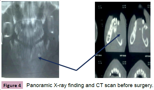
The Panoramic radiograph showed cystic radiolucent defect of the horizontal part of the left mandible; this oval-shaped lesion measured out 4 cm × 3 cm was overlying beneath incisor and canine group of teeth with a tooth bud being expelled throughout vestibular border of the mandible. The CT scanner noted multilocular enormous tumour of the mandible, blowing its cortical in some places therefore evoking an ameloblastoma.
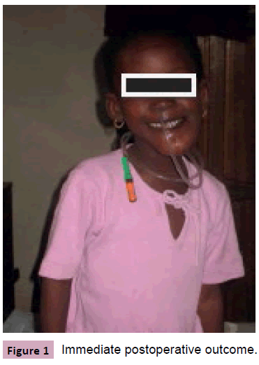
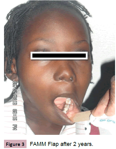
The child underwent surgery within vestibular sulcus approach by a longitudinal incision lying 5 mm beneath attached gingiva. Thus a periosteal sharp dissection was performed locating the left mentum foramen and exposing the mandible horizontal part. A shutter created in the lateral side of the bone discovered an enormous tumour with 6 cm main axis length, fit into its capsule and blowing the mandible wall. A conservative surgery was decided consisting in tumour excision as a whole followed by curettage and aspiration of the bone crater within muscularmucosal suture. There were persistent bone tissue defects restored on the 24th post-operative day using a homolateral inferior pedicled FAMM Flap.
The histological examination confirmed multicystic ameloblastoma diagnosis and there wasn’t distant recurrence after four years of follow-up.
Discussion
Ameloblastoma is a very rare tumour before 10 y of age and exceptional in early childhood before 6 y. All the authors report a high frequency in the young adult with 90% of location on the mandible [3,4]. The lesions such as dental avulsion, infections like periodontitis, pericementitis, cellulitis, have been incriminated by some authors to further epithelial stump cells proliferation of dental organ [5]. None of those factors was found in our patient.
The study has noticed the latest of consultation with a long period of five months before diagnosis and surgery. This situation could be explained by the asymptomatic long run of the tumour, low standard of living and lack of qualified maxillofacial surgery fit to the child in our regions. ZAHRA in MOROCO found a mean period of 25 months before surgery in her thesis about 10 cases with only one child aged 13 y [6].
The clinical feature was resumed by an isolated voluminous mass progressively setting and blowing the left mandible. Most of the authors have report painless and asymptomatic aspect of the tumour in its growth [7]. Some abnormalities associated to the swelling such as gingival bleeding, dental mobility or malocclusion has been reported, however [8].
Panoramic X-ray realized in our girl discovered cystic radiolucent defect off the left mandible while Computed Tomography demonstrated multilocular lesion blowing bone’s cortical. The imaging typical feature of ameloblastoma is described either as multilocular with thin grooves taking on the appearance of ‘‘soap bubbles’’, or as unilocular simulating odontogenic cyst [9]. The CT-scanner and Panoramic radiograph in our patient were very suggestive of ameloblastoma but, authors as LEZY stress on the interest of MRI in tumour extension assessment [10].
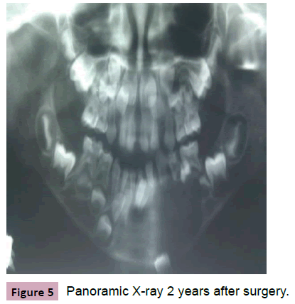
Conservative surgery was performed in this case followed by reconstruction of hard loss tissue using a FAMM’s flap. This therapeutic option was guided by the young age and aesthetic concern for our girl. MOSQUEDA has proposed mandible segmental ablation with bone graft reconstruction [11]. We believe as well as ECKARDT that radical surgery presents specific complicated problems of growth in early childhood [12]. BINGER in Germany has justified indication of conservative surgery by the low risk of recurrence in the child compare to the adult [4]. The particularity in our treatment is the use of FAMM flap in bone covering (Figures 1-5).
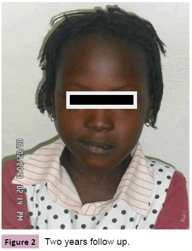
The follow-up has showed after four years no recurrence with functional and aesthetic good results. Analysis of cystic ameloblastoma enucleation surgery world reviewed in 20 children after 5 y follow-up found recurrence in around 40% of cases [3].
Conclusion
Ameloblastoma in early childhood is very rare and seems different from adult one with a high rate of unicystic type. The diagnosis is evoked on CT scanner view and confirmed after histological examination. Its conservative surgery is justified on one hand by the low risk of recurrence comparatively to adult one and on the other hand by specific problems of growth in bone resection option. This study has stressed on very young age of our case with giant aspect of tumor at the diagnosis time and the recourse of FAMM’s flap for filling bone loss.
References
- Adou A, Souaga K, Konan E, Assa A, Angoh Y (2002)Améloblastome du sinus maxillaire à propos d’un cas. Oral Surg Oral Pathol 93: 13-20
- Vallicioni J, Loum B, Dassonville O, Poissonet G, Ettore F, et al.(2007) Les améloblastomes: Annales d’Oto-Rhino-Laryngologie et de chirurgie cervicofaciale 124: 166-171.
- Ord RA, Blanchaert RH, Nikitakis NG, Sauk JJ (2002)Ameloblastoma in children. J Oral maxillofacial Surgery 60: 762-770.
- Binger T, Jundt G (1988) Single cystic ameloblastoma at an early case: 2case report. Mund Kiefer Gesichtschir 4: 213-215.
- Delaire J, Billet I, Lumineau JP, Schmidt J (1980) Le traitement chirurgical des grands kystes des maxillaires. RevStomatolchirmaxillofac81: 3-9.
- Zahra F (2011)Ameloblastome mandibulaire: étude rétrospective à propos de 10cas. Thèse Fès Maroc: n°135/11.
- Gadegbeku AS, Creuzoit GBE, Adou A, Angoh Y, Marega FB (1994) L’ameloblastome en milieu africain. RevStomatolChirMaxillofac95: 70-73.
- Jeblaoui Y, Ben neji N, Haddad S, Ouertatani L, Hchicha S (2007) Algorithme de prise en charge des améloblastomes en Tunisie. Revue de Stomatologie et de Chirurgie Maxillofaciale 108: 419-423 Masson, Paris, France.
- Shame E, Leong J, Maher R, Schenberg M, Leung M, et al. (2009) Mandibular ameloblastoma: clinical experience and literature review. ANZ journal of Surgery79: 739-744.
- Lezy J P, Princ G (1987) Abrégés de stomatologie et pathologie maxillo-faciale. 2èmeéds, 128-129.
- Mosqueda-Taylor A, Carlos-Bregni R, Ramirez-Amador V, Palma-Guzman JM, Esquivel-Bonilla D, et al. (2002)Odonto-ameloblastoma: clinico-pathologic study of three cases and critical review of the literature. Oral Oncol 38:800-805.
- Eckardt A, Swennen G, Barth EL, Brachvogel P (2006) Long-term results after mandibular continuity resection in infancy: the role of autogenous rib grafts for mandibular restoration. J Craniofac Surg. 17: 255-260.
Open Access Journals
- Aquaculture & Veterinary Science
- Chemistry & Chemical Sciences
- Clinical Sciences
- Engineering
- General Science
- Genetics & Molecular Biology
- Health Care & Nursing
- Immunology & Microbiology
- Materials Science
- Mathematics & Physics
- Medical Sciences
- Neurology & Psychiatry
- Oncology & Cancer Science
- Pharmaceutical Sciences
